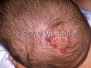
PHACE syndrome in Infant/Neonate
Alerts and Notices
Important News & Links
Synopsis

PHACE(S) syndrome (posterior fossa malformations, hemangioma, arterial anomalies, coarctation of the aorta and cardiac defects, eye / endocrine abnormalities, sternal defects) represents a neurocutaneous entity most often recognized by the presence of a large (> 5 cm) hemangioma occupying a facial segment. The hemangioma may occur on the face, scalp, or cervical region and adjacent upper extremity. About 90% of patients have infantile hemangiomas located on the head; 70% of those affected will have only one other extracutaneous manifestation of the syndrome. The most common extracutaneous finding is an arterial anomaly. Dysgenesis, anomalous origin, and narrowing, respectively, are the most frequent arterial anomalies, the presence of which confers risk for hypoperfusion, thrombosis, and cerebral infarction, eventuating in seizures and developmental delay. Examples include arterial aneurysms, anomalous branching of the carotid artery, or coarctation of the aorta. Other potentially associated features include Dandy-Walker malformation (ie, cerebellar vermis agenesis or hypoplasia, cystic dilatation of the fourth ventricle, and enlarged posterior fossa) and cardiac septal defects. Eye abnormalities include congenital cataracts, increased vascularity, microphthalmia, and hypoplasia of the optic nerve. When sternal defects such as clefting and supraumbilical abdominal raphe are present, the constellation of findings is termed PHACES syndrome instead of PHACE syndrome.
PHACE syndrome is thought to arise from a developmental error occurring during the early vasculogenesis of the first trimester. Ninety percent of those affected are female, and no familial tendency is known. PHACE syndrome can be diagnosed by the presence of the large segmental hemangioma and 1 major or 2 minor criteria; possible PHACE syndrome is ascertained when the hemangioma is only accompanied by 1 minor criterion (see References).
There appears to be some correlation between the facial segment occupied by the hemangioma and the underlying vascular / structural malformations. For instance, hemangiomas involving segments 1 and 4, the frontotemporal and frontonasal areas, have a higher risk for cerebrovascular and ophthalmologic defects. Hemangiomas involving mandibular segment 3 have an increased incidence of midline and cardiovascular defects, in addition to the laryngeal involvement associated with this area. Interestingly, if the hemangioma is unilateral, underlying structural anomalies of the brain tend to occur on the same side, presumably from a contiguous defect.
Other emerging associations that may present beyond infancy include headaches, speech delay, auditory anomalies, dysphagia, delayed puberty, and dental anomalies.
Codes
ICD10CM:
Q87.89 – Other specified congenital malformation syndromes, not elsewhere classified
SNOMEDCT:
78572006 – Neurocutaneous syndrome
Q87.89 – Other specified congenital malformation syndromes, not elsewhere classified
SNOMEDCT:
78572006 – Neurocutaneous syndrome
Look For
Subscription Required
Diagnostic Pearls
Subscription Required
Differential Diagnosis & Pitfalls

To perform a comparison, select diagnoses from the classic differential
Subscription Required
Best Tests
Subscription Required
Management Pearls
Subscription Required
Therapy
Subscription Required
References
Subscription Required
Last Reviewed:05/10/2020
Last Updated:01/20/2022
Last Updated:01/20/2022

