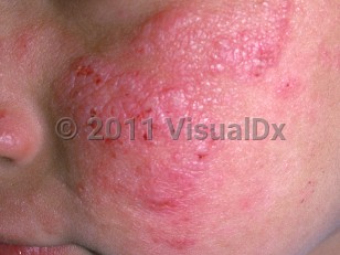
Actinic prurigo in Child
Alerts and Notices
Important News & Links
Synopsis

Actinic prurigo (AP), also called Hutchinson's summer prurigo or hydroa aestivale, is a photoinduced disorder triggered by exposure to ultraviolet (UV) radiation. It presents with intensely pruritic papules, plaques, or nodules that are typically excoriated and may scar. Secondary lichenification or eczematization may also occur.
AP is a familial dermatosis affecting primarily Native Americans and persons of mixed ancestry (Mestizo) located in the Americas, mostly Central and South America. It seems to be more prevalent in dryer, warm climates above 1000 meters in altitude. There is some evidence that cohabitation with domestic and farm animals and exposure to wood smoke are separate risk factors. It has also been described in White and Asian populations. AP usually presents in childhood, with a mean age of onset prior to 10 years. There is a female preponderance of approximately 4:1 in the Americas, whereas studies indicate a higher proportion of adult-onset cases along with a male preponderance in Asian populations.
There is a strong association with the HLA-DR4 antigen (90% of cases), suggesting an autoimmune basis for AP, the most specific subtype being DRB1*0407 (60% of cases), with DRB1*0401 next most prevalent at up to 20% of cases.
AP is initiated by exposure to UV radiation, with the UVA spectrum more strongly implicated than UVB. Current evidence, including findings of eosinophils and mast cells in the dermal infiltrate of mucosal and/or skin lesions along with high levels of immunoglobulin E (IgE) correlating with disease severity, suggests that UV exposure triggers a delayed type IVb hypersensitivity response to an as yet unclassified epidermal antigen. There is an overproduction of tumor necrosis factor alpha (TNF-α) in the suprabasilar keratinocyte layer that leads to production of clinical lesions.
The hallmark symptoms of AP are intense pruritus of involved skin, with the majority of patients also experiencing oral (lip) tingling and pain. Additional ocular symptoms include photophobia and increased lacrimation.
Clinically, there are excoriated, pruriginous papules and nodules in photoexposed areas. Cheilitis and/or conjunctivitis may occur in up to 50% of patients.
In more temperate latitudes, the disease is more seasonal, with exacerbations in the spring and persisting through summer. Even in these climates, portions of the rash may persist through winter. In the more equatorial latitudes, AP is a year-round condition.
Many of those afflicted as children will spontaneously remit by late adolescence, but a chronic course persisting into adulthood is common. Cases with adult onset tend to be more chronic.
Codes
ICD10CM:
L56.4 – Polymorphous light eruption
SNOMEDCT:
201015007 – Actinic prurigo
L56.4 – Polymorphous light eruption
SNOMEDCT:
201015007 – Actinic prurigo
Look For
Subscription Required
Diagnostic Pearls
Subscription Required
Differential Diagnosis & Pitfalls

To perform a comparison, select diagnoses from the classic differential
Subscription Required
Best Tests
Subscription Required
Management Pearls
Subscription Required
Therapy
Subscription Required
References
Subscription Required
Last Reviewed:01/18/2021
Last Updated:01/18/2021
Last Updated:01/18/2021

