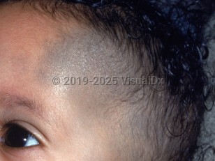
Nevus of Ota - External and Internal Eye
See also in: OverviewSynopsis

The visible pigment arises from an increased number of dermal dendritic melanocytes, and the blue and gray color seen is secondary to the Tyndall effect. Nevus of Ota occurs with greater frequency in patients with Asian or African ancestry. The mutations driving nevi of Ota may result in clinical manifestations as soon as early infancy. Darkening and extension may occur with ultraviolet (UV) radiation exposure and with hormonal influences around puberty.
Malignant transformation is very rare, but melanoma has been reported, possibly due to a second-hit mutation in the same molecular pathway. Importantly, at least 2 pediatric patients (aged 16 years and 18 years) and a few more patients aged younger than 30 years with nevi of Ota have been reported as having developed facial melanoma. At least 18 cases of central nervous system melanoma have also been reported in patients with nevi of Ota; the youngest patient was 21 years old. Ipsilateral glaucoma is a contiguous potential association in 10% of patients with nevi of Ota. The mutations associated with nevi of Ota have also been found in uveal and remote cutaneous melanomas, which may occur in 1:400 cases. Two 24-year-old patients with nevi of Ota developed cutaneous melanomas with local and distant metastases. A 29-year-old patient with a nevus of Ota developed a retro-orbital ocular melanoma that metastasized to the brain. Meningeal melanocytomas have also been associated with nevi of Ota; they are usually located ipsilateral to the nevus.
Nevus of Ota may be a feature of phakomatosis pigmentovascularis, which has also been determined to feature GNAQ / GNA11 mutations in some cases.
Related topic: choroidal melanoma
Codes
D22.39 – Melanocytic nevi of other parts of face
SNOMEDCT:
254817005 – Nevus of Ota
Look For
Subscription Required
Diagnostic Pearls
Subscription Required
Differential Diagnosis & Pitfalls

Subscription Required
Best Tests
Subscription Required
Management Pearls
Subscription Required
Therapy
Subscription Required
References
Subscription Required
Last Updated:08/11/2021
 Patient Information for Nevus of Ota - External and Internal Eye
- Improve treatment compliance
- Reduce after-hours questions
- Increase patient engagement and satisfaction
- Written in clear, easy-to-understand language. No confusing jargon.
- Available in English and Spanish
- Print out or email directly to your patient


