Common acquired nevus - External and Internal Eye
See also in: Overview,Hair and Scalp,Oral Mucosal LesionAlerts and Notices
Important News & Links
Synopsis

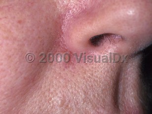
The skin of the eyelid may develop pigmented nevi; their appearance, classification, and malignant potential are the same as elsewhere on the skin. Importantly, acquired pigmented lesions after age 35–40 should be considered more suspicious of being something other than a benign nevus.
The conjunctiva and episclera may also develop nevi (which may also actually be congenital with delayed recognition); as in skin, these may be histologically junctional, compound, or deep. The nevus is the most common pigmented lesion of the conjunctiva, generally becoming apparent in the first or second decade of life and over time becoming more pigmented.
The conjunctiva and episclera may also develop nevi (which may also actually be congenital with delayed recognition); as in skin, these may be histologically junctional, compound, or deep. The nevus is the most common pigmented lesion of the conjunctiva, generally becoming apparent in the first or second decade of life and over time becoming more pigmented.
Codes
ICD10CM:
D22.9 – Melanocytic nevi, unspecified
SNOMEDCT:
400096001 – Melanocytic nevus
D22.9 – Melanocytic nevi, unspecified
SNOMEDCT:
400096001 – Melanocytic nevus
Look For
Subscription Required
Diagnostic Pearls
Subscription Required
Differential Diagnosis & Pitfalls

To perform a comparison, select diagnoses from the classic differential
Subscription Required
Best Tests
Subscription Required
Management Pearls
Subscription Required
Therapy
Subscription Required
References
Subscription Required
Last Updated:02/25/2010
 Patient Information for Common acquired nevus - External and Internal Eye
Premium Feature
VisualDx Patient Handouts
Available in the Elite package
- Improve treatment compliance
- Reduce after-hours questions
- Increase patient engagement and satisfaction
- Written in clear, easy-to-understand language. No confusing jargon.
- Available in English and Spanish
- Print out or email directly to your patient
Upgrade Today

Common acquired nevus - External and Internal Eye
See also in: Overview,Hair and Scalp,Oral Mucosal Lesion

