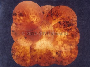
Retinitis pigmentosa - External and Internal Eye
Synopsis

The clinical picture may occur in isolation or as part of numerous syndromic conditions. These include abetalipoproteinemia, Usher syndrome, Biemond syndrome type 2, Jalili syndrome, Senior-Løken syndrome, Bardet-Biedl syndromes, disorders of glycosylation, Kearns-Sayre syndrome, gyrate atrophy, and neurodegeneration with brain iron accumulation (formerly Hallervorden-Spatz disease).
There is no known risk factor to RP. Many gene mutations are associated with it, the most common being the rhodopsin gene (RHO). RP can occur as a sporadic mutation or be autosomal dominant, recessive, or X-linked. Most autosomal recessive cases are associated with other systemic disorders. Patients can undergo genetic testing to see which gene mutations are associated with which disease. Mutation in RPE65 is particularly important as that mutation is the only one approved for genetic therapy.
True RP is a nonsyndromic ocular disease with a characteristic clinical picture as described here, while many of the pigmentary changes in the fundi of individuals with associated syndromes are more variable and should be called "pigmentary retinopathy" instead.
Codes
H35.52 – Pigmentary retinal dystrophy
SNOMEDCT:
28835009 – Retinitis pigmentosa
Look For
Subscription Required
Diagnostic Pearls
Subscription Required
Differential Diagnosis & Pitfalls

Subscription Required
Best Tests
Subscription Required
Management Pearls
Subscription Required
Therapy
Subscription Required
References
Subscription Required
Last Updated:01/23/2022

