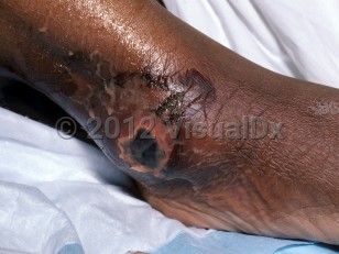
Necrotizing fasciitis - External and Internal Eye
See also in: Overview,Cellulitis DDxAlerts and Notices
Important News & Links
Synopsis

If you are considering this diagnosis, stop reading this and contact a surgeon now. The mortality of necrotizing fasciitis is high. Treatment includes broad-spectrum intravenous (IV) antibiotics and immediate surgical debridement of infected and devitalized tissue.
However, due to the rich vascular supply and thin skin of the eyelids, necrotizing fasciitis limited to this area is often treated with a more conservative surgical approach to preserve eyelid function and appearance. The prognosis is often good for patients with necrotizing fasciitis limited to the eyelids.
Diagnosis Overview:
Necrotizing fasciitis is a deep and often devastating bacterial infection that tracks along fascial planes and expands well beyond any outward cutaneous signs of infection (eg, erythema). It may be classified as polymicrobial (type 1) or monomicrobial (type 2). Type 1 infections are caused by aerobic and anaerobic organisms and generally affect hosts who are immunocompromised, those with underlying illness (such as diabetes mellitus), and elderly patients. Type 2 infections are most commonly caused by Streptococcus pyogenes, although they can be caused by methicillin-resistant Staphylococcus aureus (MRSA); they can occur in healthy individuals with no past medical history.
Necrotizing fasciitis can occur without a clear portal of entry, although predisposing risk factors include major penetrating trauma (eg, crush injury, deep penetrating wound), minor nonpenetrating trauma (eg, muscle strain, sprain, or contusion), and breaches in the skin and mucosa (eg, lacerations, varicella vesicles, insect bites, and surgical wounds). Vibrio vulnificus is associated with necrotizing fasciitis infections in cirrhotic patients with exposure to ingestion of raw oysters and with exposure of lacerations to salt water. Aeromonas hydrophila is part of the Vibrionaceae family and can cause necrotizing fasciitis in both immunocompromised and immunocompetent patients. Unlike V vulnificus sepsis, where exposure is usually to seawater, in A hydrophila infection, contact with brackish water, soil, wood, or dirty ditches is typically the common exposure. Infections can follow any trauma, fracture, or injury where there was exposure to fresh water. Infection has also occurred in the setting of debris or floodwater after a hurricane. Both V vulnificus and Aeromonas infections can present with hemorrhagic bullae, purpura, and skin necrosis. Aeromonas hydrophila infection, in contrast to infection with V vulnificus, is marked by more myonecrosis and a distinctive foul odor when the wound is debrided. Most patients with A hydrophila had exposure to wet soil or dirty ditches.
Patients with necrotizing fasciitis are acutely ill. They are often thought to have cellulitis that is not responding to standard antibiotic therapy. There is commonly a paucity of cutaneous findings in the early course of the disease. Pain is out of proportion to physical findings. There may be associated skin necrosis and bullae formation. While necrotizing fasciitis most commonly involves the lower extremities, other sites may also be involved. Signs of systemic illness such as fever, lethargy, hypotension, and tachycardia are present; these may progress to multiorgan failure.
Note: In December 2022, the Centers for Disease Control and Prevention (CDC) issued a Health Alert Network (HAN) Health Advisory to notify clinicians and public health authorities of an increase in pediatric invasive group A streptococcal (iGAS) infections in several states. During 2023, there has also been an increase in iGAS infections among persons aged 65 and older. Other groups at higher risk for iGAS infections include American Indian and Alaska Native populations; residents of long-term care facilities; people with underlying medical conditions, including diabetes, malignancy, immunosuppression, chronic kidney disease, cardiac disease, or respiratory disease, wounds, and skin disease; people who inject drugs; and people who are experiencing homelessness. Severe outcomes of iGAS infections include necrotizing fasciitis, streptococcal toxic shock syndrome, and death.
Codes
M72.6 – Necrotizing fasciitis
SNOMEDCT:
52486002 – Necrotizing fasciitis
Look For
Subscription Required
Diagnostic Pearls
Subscription Required
Differential Diagnosis & Pitfalls

Subscription Required
Best Tests
Subscription Required
Management Pearls
Subscription Required
Therapy
Subscription Required
Drug Reaction Data
Subscription Required
References
Subscription Required
Last Updated:04/06/2023


