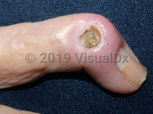
Signs and symptoms include fever, malaise, swelling, erythema, tenderness, and decreased range of motion.
Causes / typical injury mechanism: The mechanism by which osteomyelitis occurs varies with nonhematogenous versus hematogenous osteomyelitis.
- Nonhematogenous osteomyelitis occurs as a result of bacteria reaching the bones either via contact with an adjacent soft tissue infection or via direct inoculation (eg, via trauma such as open long bone fractures, surgery, or a bite wound).
- Hematogenous osteomyelitis occurs from inoculation of bone tissue with bacteria from the blood. This typically happens in the setting of bacteremia and/or endocarditis and is much more common in children.
Acute cases of osteomyelitis usually have a gradual onset over the course of several days. The following symptoms are typically present at or within the vicinity of the site of infection:
- Dull pain / tenderness that may or may not be exacerbated by movement
- Warmth
- Swelling
- Erythema
Chronic cases of osteomyelitis may have the same symptoms as acute cases, but pain, erythema, swelling, and other local symptoms might be intermittent in nature. The presence of a sinus tract, a connection between the area of osteomyelitis and the skin, is pathognomonic of chronic osteomyelitis. There may be other surrounding soft tissue insults such as soft tissue / skin ulcerations or diabetic ulcerations. Chronic osteomyelitis can be accompanied by the presence of necrotic bone (sequestrum) that may be surrounded by thickened reactive periosteal bone (involucrum).
Prevalence:
- Age – As an individual ages, the general risk for osteomyelitis increases. Additionally, the locations and pathogenesis of the disease change with age. Generally, pediatric patients will have hematogenous osteomyelitis, although hematogenous osteomyelitis of the vertebral bodies is more common in adults.
- Sex / gender – Men seem to be at higher risk.
Systemic risk factors include:
- Recent surgery
- Recent trauma / bite wounds
- Nicotine use disorder
- Obesity
- Diabetes mellitus
- Preoperative hyperglycemia
- Allergic reaction to hardware used during an orthopedic procedure
- Older age
- Immunocompromised states
- Chronic hypoxia
- Alcohol use disorder
- Malignancies
- Hepatic failure
- Renal failure
- Hypoperfusion
- Peripheral vascular disease
- Peripheral neuropathy
- Systemic infections (eg, endocarditis or bacteremia)
- Intravenous (IV) drug use
- Presence of indwelling intravascular devices
- Hemodialysis
- Sickle cell disease
- Hypoperfusion to the infected region
- Venous stasis
- Presence of surgical components
- Poorly healing soft tissue wounds
The most common causative agent by far of osteomyelitis is Staphylococcus aureus (including methicillin-resistant S aureus [MRSA]). Other common causes include coagulase negative staphylococci (eg, Staphylococcus epidermidis) and aerobic gram-negative bacilli (eg, Pseudomonas aeruginosa). Corynebacteria, fungal species, and mycobacteria are less common causes.
Some species are associated with different underlying conditions:
- Staphylococcus aureus and S epidermidis are associated with IV drug users and patients who have had recent surgery and/or trauma.
- Pseudomonas aeruginosa is associated with diabetic patients.
- Salmonella spp, Streptococcus pneumoniae, and other encapsulated organisms can be associated with sickle cell disease.
- Common organisms responsible for the nosocomial setting include Enterobacteriaceae, P aeruginosa, and Candida spp.
- Fungal infections are especially associated with disseminated disease, which usually affects immunocompromised patients.
- In patients with a history of human or animal bites, consider infection with Pasteurella multocida.
Additional features of pathogenesis include superlative infective processes and accompanying inflammatory features, vascular congestion / small vessel thrombosis, necrotic bone, and formation of new bone. Furthermore, the presence of polymorphonuclear leukocytes and other inflammatory cells plays an important role in the pathogenesis of the disease.
Grade / classification system: Generally, osteomyelitis is classified by the source of the infection (ie, hematogenous or nonhematogenous) or by duration of the condition (ie, acute or chronic).
Of these classifications, osteomyelitis secondary to a contiguous infection (eg, after surgery, insertion of orthopedic hardware, or trauma) is the most common. The next most common cause is vascular insufficiency (eg, diabetic foot infections). Hematogenous origin is the least common cause of osteomyelitis, except in children in whom it is the most common form.
Classification based on the type of bone infected can also be useful, since approaches to treatment are different based on the bone type infected, specifically, long bone versus sternoclavicular versus vertebral versus pelvic versus sacral infection.
Treatment of chronic osteomyelitis includes surgical debridement of any necrotic bone or tissue followed by culture-directed antibiotic therapy. A sequestrum needs to be removed or chronic osteomyelitis may continue or reoccur. Acute osteomyelitis is frequently treatable with antibiotics alone.


 Patient Information for
